Day 1 :
Keynote Forum
Mohamed Samir Hefzy
University of Toledo, USA
Keynote: A Biologically Inspired Knee Actuator for a KAFO
Time : 10:05-10:35

Biography:
Mohamed Samir Hefzy is currently serving as Associate Dean of Graduate Studies and Research Administration of the College of Engineering and Professor of Mechanical, Industrial and Manufacturing Engineering at The University of Toledo (UT), Toledo, Ohio. He has been on the faculty of The UT since 1987. He graduated from Cairo University, Egypt, with a B.E. in Civil Engineering in 1972, and a B.Sc. in Mathematics from Ain-Shams University in 1974. He earned his M.S. in Aerospace Engineering in 1977 and his Ph.D. in Applied Mechanics in 1981, both from The University of Cincinnati. He then received training as a Postdoctoral Research Associate for two years in the Department of Orthopedic Surgery at The University of Cincinnati’s College of Medicine. In December 2003, Dr. Hefzy was elevated to the Grade of American Society of Mechanical Engineers (ASME) Fellow. He is the recipient of many awards, including the 2011 Distinguished Service Award from the ASME.
Abstract:
A person with quadriceps weakness has limited ability to perform knee extension. A knee-ankle-foot orthosis (KAFO) is a common prescription for such disability. Several types of KAFOs are currently available in the market: passive KAFOs, stance-control KAFOs and dynamic KAFOs. In passive KAFOs, the knee joint is kept locked during standing and walking. However the associated uncomfortable walking gait with high energy consumption makes these devices abandoned by patients. Stance control KAFOs block knee motion for weight bearing and allows free rotation in swing phase. However abnormal gait pattern still exists because of the locked knee joint in the stance phase. Dynamic KAFOs are developed to control both stance and swing phases. But those presently available are inconvenient to use and have complex control systems. This research is directed at using superelastic alloys to develop a biologically inspired dynamic knee actuator that can be mounted on a traditional passive KAFO. The actuator stiffness can match that of a normal knee joint during the walking gait cycle. Two superelastic actuators are used for this purpose. They are activated independently. Each actuator is developed by combining a superelastic rod and a rotary spring in series. When neither actuator is engaged, the knee joint is allowed to rotate freely. The stance actuator works only in the stance phase and the swing actuator is active for the swing phase. The conceptual design of the knee actuator was verified using numerical simulation and a prototype is being developed through additive manufacturing for confirming the concept.
Keynote Forum
Michele J Grimm,
Wayne State University, USA
Keynote: The biomechanics of neonatal brachial plexus injury
Time : 10:35-11:05

Biography:
Michele J Grimm earned her BS in Biomedical Engineering and Engineering Mechanics from Johns Hopkins and her PhD in Bioengineering from the University of Pennsylvania. She joined Wayne State in 1994 and had the opportunity to work with world leaders in injury biomechanics. In 1997, she began collaborating with an obstetrician on a model of shoulder dystocia and NBPP. She has since become a recognized expert in this area and was the only engineer on the American College of Obstetricians & Gynecologists working group on NBPP. She is a Fellow of ASME and a past chair of the Bioengineering Division of ASME.
Abstract:
Approximately 1 in 1000 infants is noted to have a brachial plexus palsy at the time of birth – resulting in paralysis of the arm. About 10% of these injuries are “permanent” – with residual paralysis after 1 year-of-age. For over 100 years, efforts have been made to understand the mechanism of neonatal brachial plexus palsy (NBPP) and reduce its incidence. Due to the relatively rare nature of the injury, and the sensitivity of studies involving pregnant women and infants, typical experimental methods in injury biomechanics have not been appropriate for NBPP. In the past 15 years, modeling techniques – both computer physical models – have been developed to gain greater insight into NBPP injury mechanisms. But the development of models that cannot be fully validated presents its own challenges. Both computer and physical models have demonstrated that significant stretch of the brachial plexus occurs both as a result of the natural, maternal forces of delivery (uterine contractions and maternal pushing) and any assistive traction applied by the clinician. Available data indicates that stretch due to maternal forces alone is sufficient to cause permanent NBPP in some infants. Currently, there is no way to characterize clinically which individuals will be more susceptible to nerve injury than others. This presentation will review the current state of the art with respect to models of NBPP, with particular focus on the development of computer models, in addition to the current data regarding nerve injury thresholds. The gaps in knowledge that deserve to be addressed will be identified.
Keynote Forum
Lisa A Ferrara
OrthoKinetic Technologies, LLC, USA
Keynote: Innovative biomaterials and surface technologies for medical devices: Observations of mechanotransduction and biologic induction
Time : 11:25-11:55

Biography:
Dr. Lisa Ferrara has been faculty at two prestigious academic medical centers and served as the director of the musculoskeletal research facilities. She has received numerous accolades, was involved with the Medical Device Advisory Committee to the FDA, and has provided consulting services about spinal disorders for ABC News. Dr. Ferrara is widely published, provides frequent lectures, serves on multiple scientific and medical advisory boards, and has recently been appointed as a board member to the Advisory Committee for Biotechnology in Southeastern North Carolina.\\r\\n\\r\\nDr. Ferrara previously served as the Director of the Spine Research Laboratory in the Department of Neurosurgery and Orthopedics at The Cleveland Clinic with a research focus on musculoskeletal biomechanics and the development of implantable MEMS sensors for various biomedical applications. She was awarded the Who’s Who Award in Technology in 1999, the NASS Award for Outstanding Research in 1995, and is the recent recipient of the Healthcare Entrepreneur of the Year Award for Coastal North Carolina. \\r\\n
Abstract:
Over multiple decades, orthopedic medical devices have been fabricated from various materials including metals, polymers, ceramics, and allograft tissue. Each material has intrinsic properties that possess certain mechanical characteristics designed for specific anatomical regions and biomechanical parameters. Numerous challenges still exist with the long-term success of many orthopedic devices, yet as innovation continues to progress rapidly, novel materials and surface geometries offer the potential to optimize interface mechanics for improved osseointegration and performance. Therefore, medical devices must be designed with an understanding of the biological and biomechanical principles at the macro, micro, and nano level to offer improved design and implant function. Optimized surface geometries and novel biomaterials have the potential to improve mechanotransduction, while providing greater biological compatibility and biomechanical synergy for long term implantation. These new technologies potentially possess antibiotic/anti-infective properties, micro/nano-surface technologies for improved osseointegration, and controlled surface structures for directed cellular differentiation. Surface geometries and multidimensional load bearing scaffolds have been incorporated into orthopedic implant designs to provide greater contact surface areas at the tissue and bone interface and contribute to improved performance for long term stabilization and immobilization of the tissue. Additionally, the advent of new manufacturing technologies can develop intricate structural scaffolds and nanosurfaces that provide controlled and predictable mechanical stimuli to the cell, where mechanotransduction can be optimized to improve implant function. Therefore, the objective of this work will be to present an overview of a few of these novel technologies, and to present the effects of these novel biomaterials, surface technologies, and structural designs on mechanotransduction and tissue healing.
- Nano-Mechanical Implant Design
Session Introduction
Mark Pitkin
Tufts University School of Medicine, USA
Title: Addressing anisotropy of tissues regeneration in the implants design
Time : 12:00-12:25
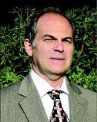
Biography:
Mark Pitkin has completed his PhD in Biomechanics at the Central Institute of Prosthetics Research in Moscow, Russia. He is Professor of Physical Medicine and Rehabilitation at the Tufts University School of Medicine, Boston, MA.
Abstract:
Anisotropy is a fundamental characteristic of tissues regeneration replicating those in the tissues development. If design of an implant is not addressing it properly, the long-term bond between implant and the hosting tissues can-not be sustainable neither in total joint replacement, nor in direct skeletal attachment (DSA) of limb prostheses. Advantages in utilizing anisotropy of regeneration were demonstrated by preferred properties and orientation of the components of the Skin and Bone Integrated Pylon (SBIP) developed by Poly-Orth International, Sharon, MA. Besides optimizing porosity, pores size, particle size and volume fraction, we improved skin-implant and bone-implant interface via preferred orientation of the pylons’ parts. Skin-implant interface: The SBIP is deeply porous perpendicular to the implant’s longitudinal axis and also has perforations in the solid enforcing inserts. Skin cells can therefore penetrate the structure and grow throughout the entire volume of the implant in the “natural” anisotropic direction of regeneration. That creates a skin seal, thus addressing the principal failure modes in existing percutaneous devices: skin regression, marsupialization, permigration, and avulsion. Bone-implant interface: Bone loss after implantation to the marrow canal is observed, and is caused by shield stresses. A variant of the SBIP and a new method of fixation are addressing this problem. Our SBIP-F pylon has side fins that are inserted into precut slots inside the cortical bone walls. The cut out bone activates an Ilizarov type distractional osteogenesis in which the regenerated bone has greater strength than the original bone.
Yoichi Aota
Yokohama Brain and Spine Center, Japan
Title: Entrapment of the cluneal nerves as a forgotten cause of low back pain and leg symptoms
Time : 12:25-12:50
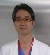
Biography:
Yoichi Aota has completed his PhD from Yokohama City University and Postdoctoral studies from Rush-Presbyterian - St. Luke’s Medical Center, Chicago. He is the Director of department of spine & spinal cord surgery of Yokohama Brain and Spine Center and a visiting Professor of Yokohama City University and Tokyo Medical University. He has published more than 40 papers in reputed journals and has been serving as an Editorial Board Member of repute.
Abstract:
The causes of low back pain (LBP) can be complex and it is often difficult to get an accurate diagnosis. The concept of a relationship between the cluneal nerve and LBP is not new. Following reports by Maigne et al. in 1989 describing that the most medial branch of the superior cluneal nerve (SCN) may become entrapped where the nerve passes through the fascia over the iliac crest, surgeries were undertaken for irritative SCN neuropathy to open the fascial orifice with successful outcomes. Recently, clunealgia has become known as an under-diagnosed cause for chronic LBP or leg pain. We reported that patients with SCN disorders comprised 12% of all patients presenting with a chief complaint of LBP and/or leg symptoms in our clinic and approximately 50% of SCN disorder patients had leg pain and/or tingling. There are several anatomical variations in the running courses of the SCN. On the other hand, entrapment of the middle cluneal nerve (MCN) within the long posterior sacroiliac ligament (LPSL) has been sporadically suggested as a potential cause of LBP and peripartum pelvic pain. In my experience, SCN entrapment is often associated with MCN entrapment. MCN entrapment under the LPSL is a potentially under-diagnosed cause of chronic LBP. Knowledge of this clinical entity would avoid unnecessary spinal surgeries and sacroiliac joint fusion. SCN and/or MCN blocks are useful not only for obtaining pain relief, but also to confirm the diagnosis by pain relief after injection.
Boris I. Prilutsky
Georgia Institute of Technology
USA
Title: Adaptation of bone-anchored limb prosthesis for locomotor behaviors
Time : 12:50-13:15

Biography:
Boris I Prilutsky has received his PhD from Latvian Research Institute of Traumatology and Orthopedics, Riga, former USSR. He is the director of Biomechanics & Motor Control laboratory in the School of Applied Physiology at Georgia Tech, Atlanta, USA. He has published 2 books and over 60 scientific papers, has been serving as a review panel member of National Institutes of Health & National Science Foundation, USA and Natural Sciences and Engineering Research Council, Canada, and has beenan editorial board member of Scientific Reports, UK.
Abstract:
Bone-anchored limb prostheses have been utilized in Europe for over 20 years. Although this technology has many advantages over traditional socket prosthesis attachments, it has not been approved for use in US due to high risk of skin infection at the skin-implant interface. The recently developed porous titanium pylon for skin and bone integration (SBIP, Poly-Orth International, USA; Pitkin, Raykhtsaum, 2012) has demonstrated the potential for skin-bone ingrowth into the pylonthus reducing or preventing infections. Our laboratory has utilized animal models (rodents, felines) to investigate integration of SBIP pylons with residual limb after the implant is loaded during every-day prosthetic walking and standing. The animals wear a trans-tibial J-shape prosthesis over several months.Level and slope prosthetic walking is recorded using 3D motion capture and force plates. Biomechanical analysis is used to determine contributions of the prosthetic and contralateral hindlimb to joint moment, power and work production. At the end of study, animals are euthanized using deep anesthesia andlimb with implant is harvested for histological analysis. Our current results have demonstrated that animals adopt bone-anchored prostheses for standing and locomotion over several months. Although load on prostheses is reduced by approximately 30-40%, it appears caused by loss of the ankle joint, important source of mechanical energy for locomotion. Histology shows substantial skin-bone ingrowth into implant. The results strongly suggest that the SBIP can successfully be used in bone-anchored prosthesis and has the potential for reducing skin infection.
- Clinical Biomechanics
Session Introduction
Muhammed Wasif Rashid Chaudhary
Ahalia Hospital, United Arab Emirates
Title: Basics of biomechanics of the neuromusculoskeletal system and research opportunities
Time : 14:00-14:25

Biography:
Muhammed Wasif Rashid Chaudhary, MBBS, MBA, CSSGB, CTQM, CPHQ is working as Asst. Medical Director in United Arab Emirates. He has 15 years of experience in healthcare field. He had worked also as Medical Superintendent and Quality Manager 8 years in the same health care facility. His interest is in continuous quality improvement and patient safety. He is licensed general practitioner and continuing his responsibility as a physician. He is certified professional in healthcare quality (CPHQ). That credential covers field of quality, case/care/disease/utilization and risk management and emphasizes how all these programs and processes integrate into an effective system
Abstract:
Biomechanics is the study of mechanics (loads, motion, stress, and strain of solids and fluids) applied to biological systems. Musculoskeletal Biomechanics specifically focuses these methods for studies of the musculoskeletal system. This includes studies of the form and function of tissues including bone, cartilage, ligament, tendon, muscle, and nerve, at multiple scales ranging from the single cell to whole body. My presentation will provide a general overview of the biomechanical principles associated with the neuromusculoskeletal system. In particular I will review, the structure and properties of the neuromusculoskeletal system, and show how the various components of this system can be idealized and described in mathematical terms. The presentation begins with an overview of the mechanical properties of muscle, tendon, ligament, and cartilage, finishing off with a review of the ongoing research opportunities in Biomechanics, including a brief look at musculoskeletal tissue engineering, ergonomics, musculoskeletal adaptation, and tissue mechanics.
Mohamed Hamdy Doweidar
University of Zaragoza
Spain
Title: A 3D numerical model of cell differentiation and proliferation
Time : 14:25-14:50

Biography:
Mohamed Hamdy Doweidar is a Full Associate Professor at the Mechanical Engineering Department, University of Zaragoza, Spain. Besides, he is a Member of the Group of Structural Mechanics and Materials Modeling (GEMM), the Biomedical Research Networking Center in Bioengineering, Biomaterials, and Nanomedicine (CIBER-BBN), the Aragón Institute of Engineering Research (I3A), and the European Society of Biomechanics (ESB). He has participated in many national and international investigation projects. He has authored and co-authored many books and numerous articles in international journals. His investigation interests include Computational Biomechanics, Cell Simulation, Finite Element Method, Natural Element Method and Computational Fluid Dynamics.
Abstract:
Mesenchymal stem cells (MSCs) have the ability to differentiate into many cell phenotypes such as fibroblasts, chondrocytes, osteoblasts and neuronal precursors. Experimental studies have demonstrated that mechanical characteristics of the substrate, such as substrate stiffness, fluid flow and mechanical forces can govern their fate even in absence of biochemical factors. However, the signaling mechanism behind this process is not well understood. The main objective of the present work is to develop a numerical model to study the influence of substrate mechanical conditions on cell differentiation, proliferation and apoptosis. To formulate the model, a discrete finite element approach has been appreciated. For the sake of simplification, it is assumed that the cells have a spherical shape; however it is possible to consider any other cell configuration. We consider that these processes are related to the received mechanical signals by cells and its maturation time. It is assumed that the cell fate is governed by the cell internal deformation experimented during cell migration. Consistent with experimental observations, our findings indicate that within soft (0.1-1 kPa), intermediate (20-25 kPa) and hard (30-40 kPa) substrates MSCs proliferate and then differentiate into neuroblasts, chondrocytes and osteoblasts, respectively. The traction force generated by a specific cell phenotype can increase (osteoblasts and chondrocytes) or decrease (neuroblast) during differentiation. In contrast, in all cases the proliferation of a typical cell considerably increases the average cell traction force due to the cell-cell interaction. Hence, greater the substrate stiffness, the higher is the cell differentiation and proliferation rate.
Udi Sarig
Nanyang Technological University, Singapore
Title: Biophysical characterization of the recellularization effect: Restoring functional properties of ECM based constructs towared the native state
Time : 14:50-15:15

Biography:
Udi Sarig has completed his PhD from the Technion – Israel Institute of Technology (IIT) and is currently a Postdoctoral fellow at the Nanyang Technological University (NTU) in Singapore. He is a co-author of more than 20 publications in international recognized peer reviewed journals and scientific conferences, and is currently serving as a group leader in the Singapore Technion Alliance for Research and Technology (START), under the Campus for Research Excellence and Technological Enterprise (CREATE) program: The regenerative medicine initiative in cardiac restoration therapy.
Abstract:
‘Functional tissue engineering’ (FTE) – a tissue engineering (TE) subfield – employs various cellularized biomaterial scaffolds for the “engineering of load bearing tissuesâ€. To generate more biomimetic materials, various extracellular matrix (ECM) scaffolds were isolated through decellularization. However, decellularization represents a trade-off between excessive ECM damage and preservation of ECM ultrastructure and bioactivity. Indeed, vast research has identified biophysical effects of the ECM on cell survival, proliferation, migration, organization, differentiation and maturation, with clear implications for FTE. Surprisingly though, no study to date, to the best of our knowledge, provided clear methods and understanding on the reciprocal effects of cellularization on the cellularized ECM scaffolds biophysical properties, under physiological-like conditions. We hypothesized that by re-cellularizing porcine ventricular ECM (pvECM, serving as a model scaffold) some of the original myocardial tissue biophysical properties can be restored, concerning scaffolds surface and bulk modifications consequent to cellularization. We therefore performed a systematic biophysical assessment of pcECM scaffolds seeded with human mesenchymal stem cells, a common multipotent cell source in cardiac regenerative medicine. The results obtained were compared to acellular pcECM and native ventricular tissue serving as negative and positive controls, respectively. We report a new type of FTE study in which cell interactions with a composite-scaffold were evaluated from the perspective of their contribution to the construct surface (FTIR, WET-SEM) and bulk (DSC, TGA, uni-and bi-axial mechanical testing) biophysical properties. Such an approach yields important methodologies, understanding, and data serving both as a reference as well as possible ‘design criteria’ for future studies in FTE.
Yoichi Aota
Yokohama Brain and Spine Center, Japan
Title: Workshop: Entrapment of superior and middle cluneal nerves as an unknown cause of low back pain and pseudosciatica
Time : 15:35-17:10
Biography:
Yoichi Aota has completed his PhD from Yokohama City University and Postdoctoral studies from Rush-Presbyterian - St. Luke’s Medical Center, Chicago. He is the Director of department of spine & spinal cord surgery of Yokohama Brain and Spine Center and a visiting Professor of Yokohama City University and Tokyo Medical University. He has published more than 40 papers in reputed journals and has been serving as an Editorial Board Member of repute.
Abstract:
Low back pain (LBP) is one of the most common problems that most people suffer at some point in their life. There are many sources of LBP. In most LBP patients, the exact cause of LBP is not clear. Thus, one of the most difficult task with LBP is to identify the actual pain generator. Large epidemiological studies show that 20% to 37% of patients with back pain suffer from a neuropathic pain component. superior and middle cluneal nerves (SCN / MCN) entrapment must not be forgotten as cause of neuropathic LBP. Although many chiropractists, physiotherapists, and archipuncture seem to know this etiology, so far unfortunately, it is not widely recognized in orthopaedic or neurosurgeons. SCN and MCN supply the skin overlying the posteromedial area of the buttock. Previous studies illustrated that the SCN is derived from the cutaneous branches of the dorsal rami of T11-L5. In spite of more than 50 years of surgical experiences in clunealgia, information of clunealgia is limited. Entrapment of SCN/ MCN induces low back pain and leg symptoms. SCN entrapment occurs where SCNs pierce fascial attachment at posterior iliac crest. Although this clinical entity had been known as a rare cause of unilateral low back and/or buttock pain, recently, clunealgia has become known as an under-diagnosed cause for chronic LBP or leg pain. In a recent prospective study, Kuniya et al. reported that patients with SCN disorders comprised 12% of all patients presenting with a chief complaint of LBP and/or leg symptoms in their clinic and approximately 50% of SCN disorder patients had leg pain and/or tingling. The MCNs can become spontaneously entrapped where this nerve pass under the long posterior sacroiliac ligament. Clunealgia is underdiagnosed and should be considered as a potential cause of severe low-back and/or leg symptoms. The symptoms of clunealgia can be very severe and mimicked a radiculopathy and disc disorders in lumbosacral spine. Clinicians should be aware of this clinical entity and avoid unnecessary spinal surgeries and sacroiliac fusion. Techniques in SCN surgeries may differ from those in other common peripheral nerve surgeries because branches of SCN/MCN are thin and requires release in multiple branches. This workshop is to draw attention by pain clinicians in SCN/ MCN entrapment by comprehensively reviewing its historical perspective, anatomical background, clinical symptoms with respect of differential diagnosis and surgical tips.

